FIG. 1. Clinical appearance of nail changes associated with chronic kidney disease: (a) half-and-half nails characterized by a sharply demarcated distal brownish band involving approximately 50–60% of the nail plate with preserved lunulae; (b) Terry’s nails showing diffuse whitish “ground-glass” discoloration with loss of lunulae and only a narrow distal pink-brown band.

FIG. 1a. This transesophageal echocardiography image illustrates a patent foramen ovale with left-to-right shunting in a patient with infective endocarditis involving the mitral, tricuspid, and aortic valves caused by Streptococcus gallolyticus.
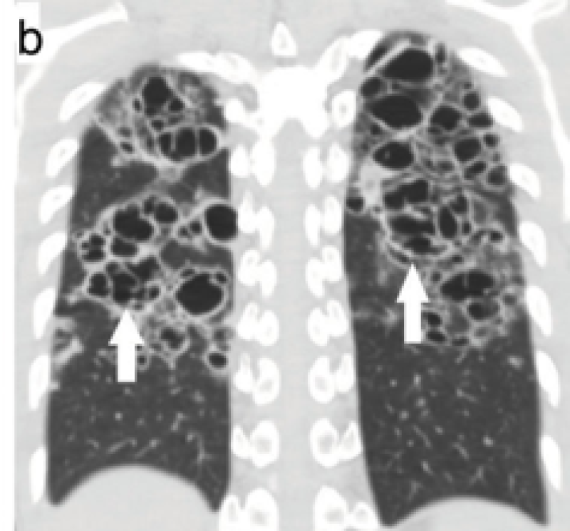
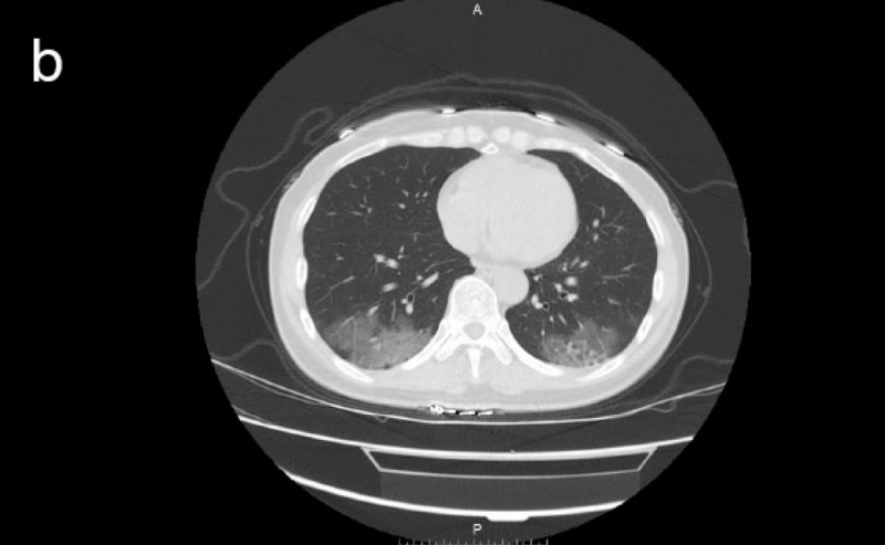
FIG. 1b. Coronal chest CT image demonstrating multiple, large cystic dilatations of the segmental bronchi consistent with extensive cystic bronchiectasis.

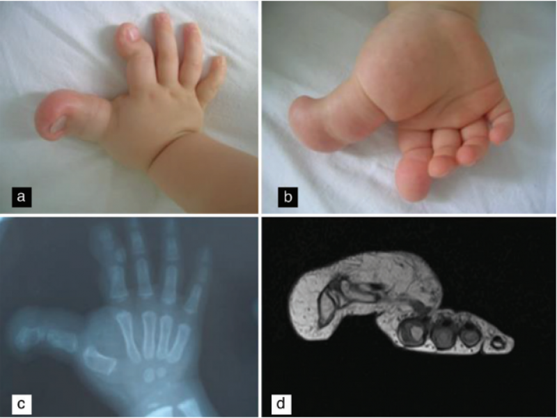
Right forearm with “fork-like” radial deviation; wrist direct radiography showing Madelung deformity
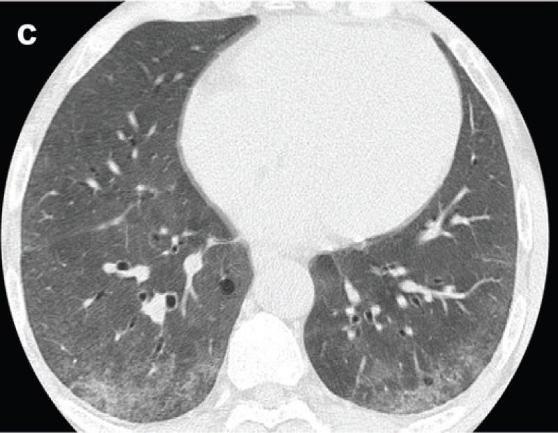
FIG. 1. Representative clinical photographs and chest CT images demonstrating findings before and after secukinumab therapy, showing regression of psoriatic skin lesions and post-treatment pulmonary changes on high-resolution CT.

3D CBCT reconstruction displaying lesion extent and bone destruction in Gorlin-Goltz Syndrome with PTCH1 Gene Mutation.
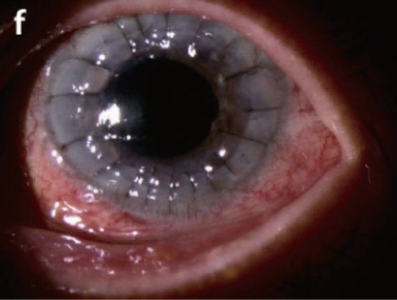
FIG. 1. (f) anterior segment photograph after the second optical penetrating keratoplasty with intrascleral intraocular lens fixation.

Computed tomography image shows a tam-o-shanter skull and generalized trabecular alteration in the skull with “cotton-wool appearance” on a Paget Disease.
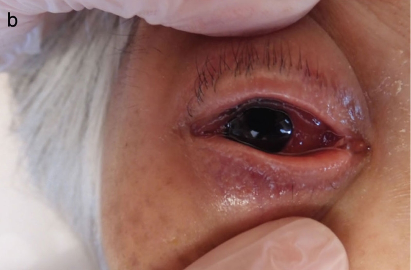
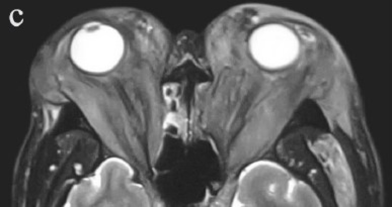
FIG. 1. Eyelid edema, chemosis and conjunctival congestion; computed tomography revealed contrast-enhanced eyelidsand extraocular muscles.

The patient was a 67-year-old man who presented with a worsening maculopapular rash on his body....

FIG. 2. (a) Pelvic magnetic resonance imaging (MRI) performed before antibiotic treatment.

FIG. 1c. Peripheral enhancement with contrast is evident on 3D-T1 contrast-enhanced imaging ….

FIG. 2. CT pulmonary angiogram 3D reconstruction reveals an aneurysmal main pulmonary artery….
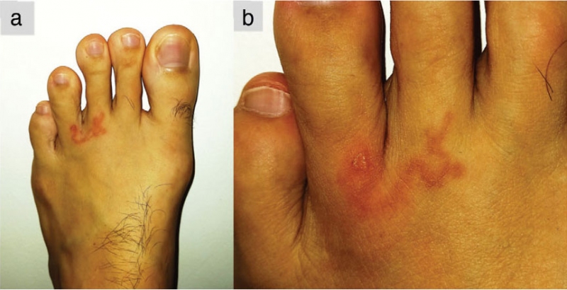
FIG. 1. 1.5-2 cm long, serpiginous, slightly-elevated, erythematous, raised tract between toes …
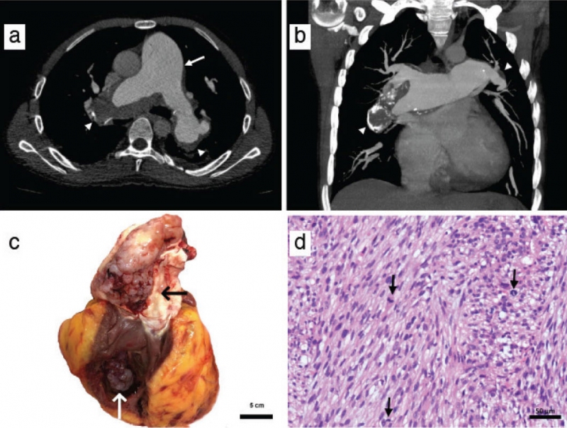
FIG. 1. (a) Axial computed tomography pulmonary angiogram (CTPA) reconstruction discovered an enlarged pulmonary trunk and filling defects partially ….
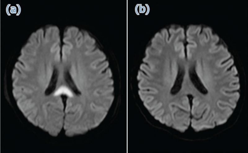
FIG. 1. Diffusion-weighted magnetic resonance imaging of the brain reveals a hyperintense signal in the splenium of the corpus callosum, without brain …
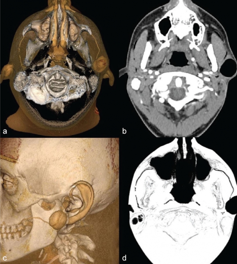
FIG. 1. Multiplanar volume-rendered , soft tissue , and minIP axial images showing the presence of air in the enlarged left parotid gland and its Stensen’s duct.…
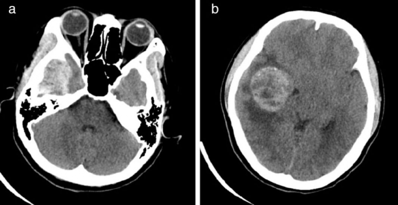
FIG. 1. CT revealed circular mixed-density shadows in the right middle cranial fossa and temporal region …
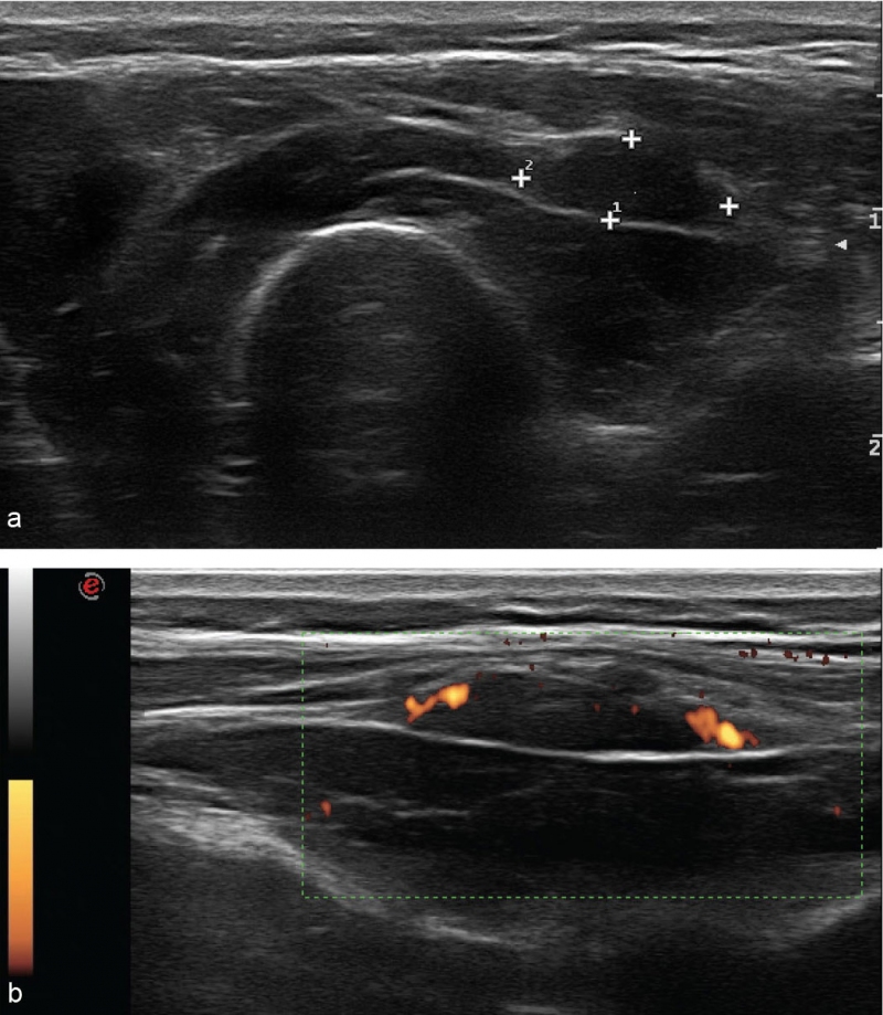
FIG. 1. a) Short-axis view of the enlarged posterior interosseous nerve within the supinator muscle…
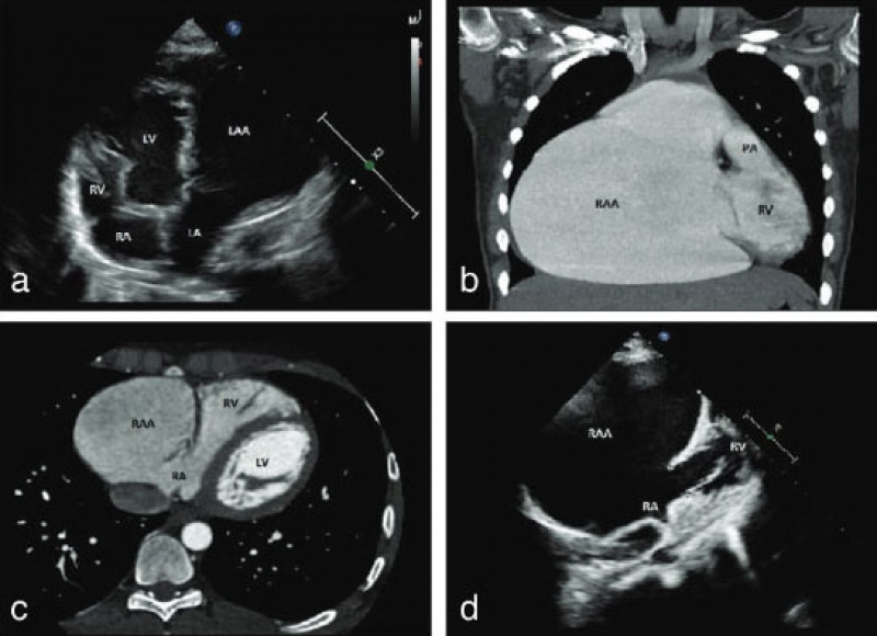
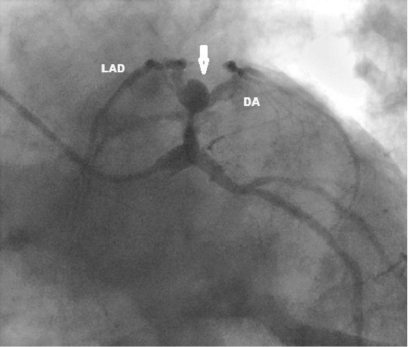
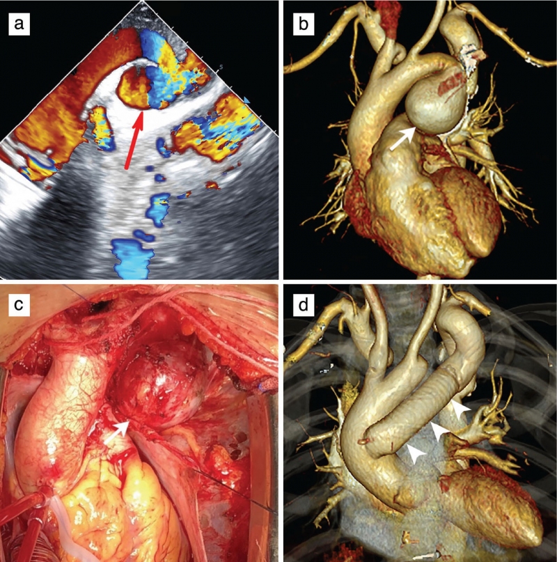
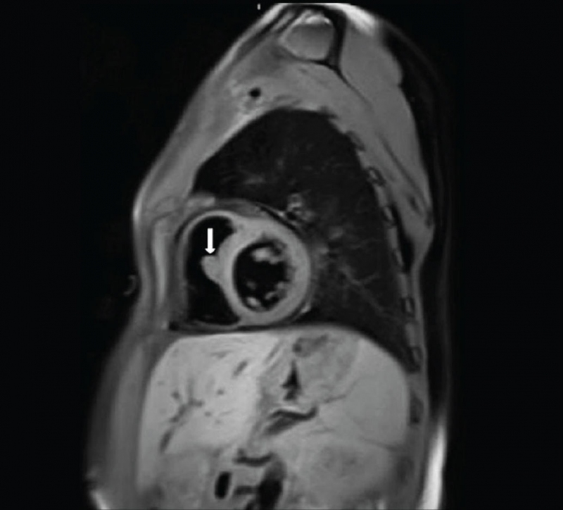
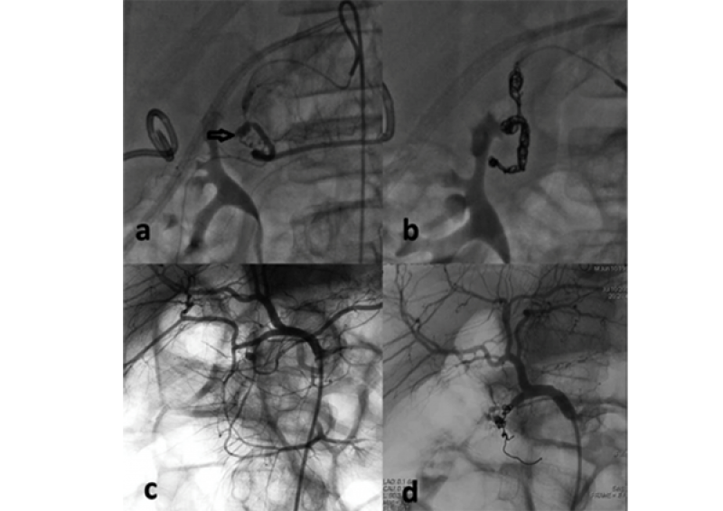
This case series describes four cases of giant aneurysms of atrial appendages (AA). Case 1: A 4.5-month-old boy was referred to us for “enlarged heart shadow on chest radiography…
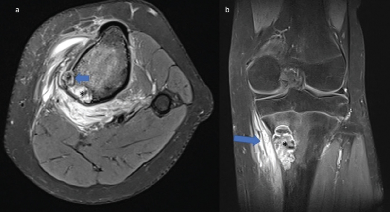
A 17-year-old male without previous trauma was admitted to the orthopedic outpatient clinic with the complaint of pain in the right knee. Plain radiography indicated a lobule-shaped, lytic lesion with a sclerotic rim in the proximal metaphyseal part of the tibia. Computed tomography revealed a soft tissue lesion …
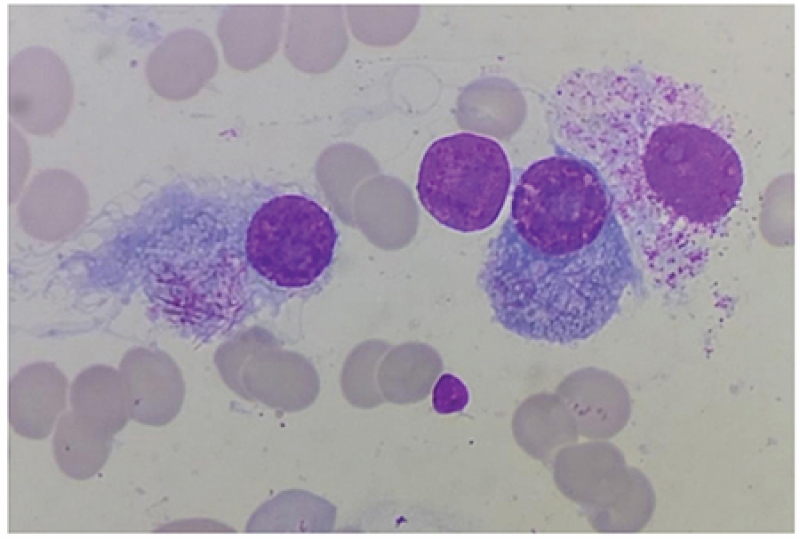
A 79-year-old man was admitted to the hematology department for an 11-month history of bone pain in the spine and pelvis, along with generalized fatigue and lethargy. He had no significant personal or family history. On general examination, he was afebrile and normotensive, with pale skin. His initial laboratory investigations …
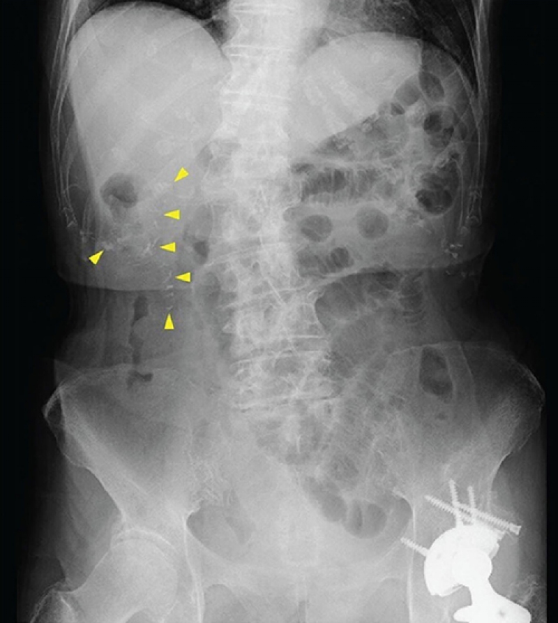
An 84-year-old Japanese woman presented to the emergency department with a 1-day history of vomiting and right lower quadrant abdominal pain. A flat film of the abdomen showed significant gaseous distention in the left quadrant and calcification in the right upper quadrant. Non-enhanced computed tomography revealed calcification of the …
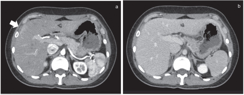
A previously healthy 28-year-old woman presented with a 2-week history of abdominal pain in the right upper quadrant and right lower quadrant. On palpation, the patient had right hypochondriac direct tenderness with positive Murphy’s sign. The patient also had hiccups, which started 2 days before consultation.
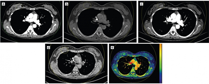
A 44-year-old woman was admitted to the emergency department with thoracic trauma to evaluate possible thoracic injury. She underwent contrast-enhanced computed tomography in a dual energy CT scanner, which is the main tomography appliance for all patients with trauma. Through virtual monoenergetic imaging, a solid lesion…
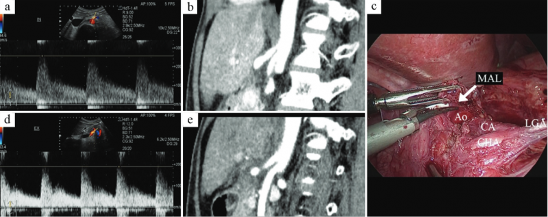
A 29-year-old female diagnosed with chronic sinusitis was referred to the ENT department because of recurrent nasal discharge without obvious incentives. Her vital signs were stable, and she had no other clinical symptoms. Two previous cardiac echocardiograms revealed no cardiovascular system diseases. Non-contrast computed tomography revealed an abnormally tortuous aortic arch with …
A 27-year-old female with depression presented to the hospital with a 4-year history of postprandial epigastric pain. One year ago, she had been diagnosed with psychogenic abdominal pain because of exhibiting no remarkable abnormalities on various examinations, including blood tests, abdominal computed tomography, and upper gastrointestinal endoscopy.

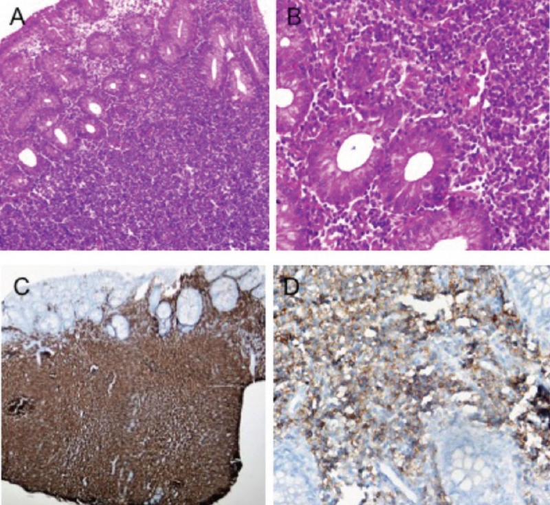
A 37-year-old woman was admitted to the hospital with a 2-month history of a growth inside the oral cavity in the upper jaw and weakness. Physical examination revealed a voluminous mass involving the left side of the maxillary gingiva. The maxillofacial computerized tomography scan confirmed the presence of a solid tissue mass at the left upper maxilla. Laboratory findings revealed a low hemoglobin level of 75 g/L, a renal impairment with creatinine level of 23 mg/L, and hypercalcemia of 191 mg/L.
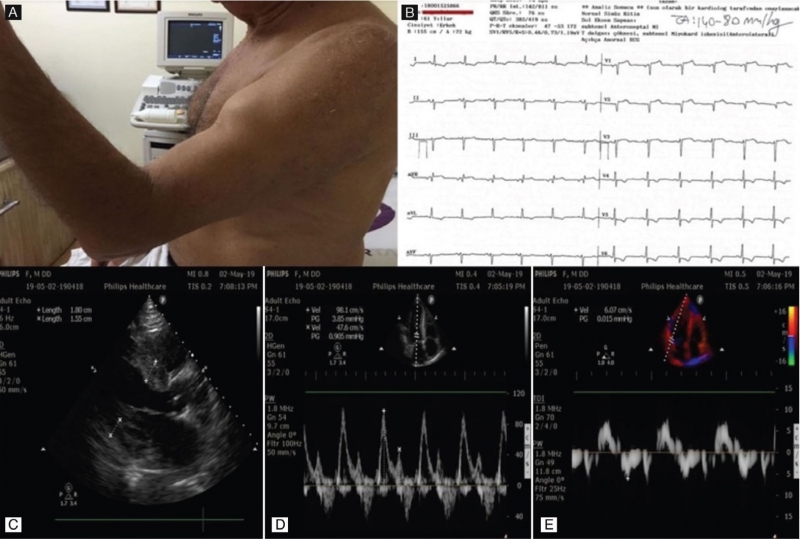
A 61-year-old man with a history of hospitalization for acute decompensated heart failure and pulmonary edema was admitted to the intensive cardiac care unit with fatigue, shortness of breath, and bilateral leg edema. Upper extremity musculoskeletal examination shows bunching of the biceps when the patient flexed his arm.
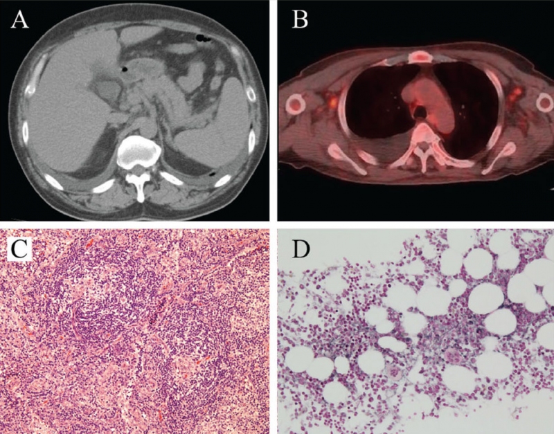
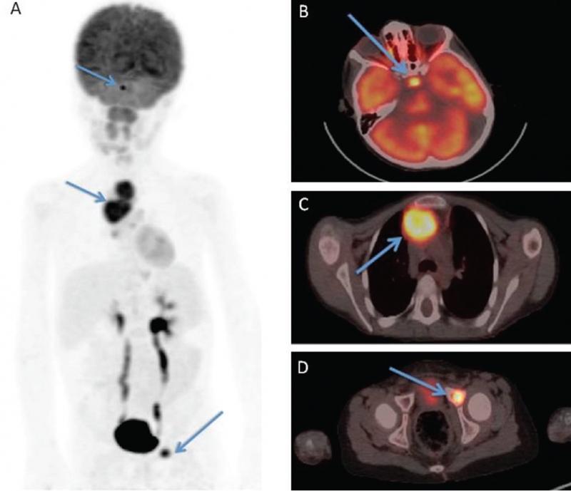
A 54-year-old male patient was admitted with a 3-day history of fever. Physical examination revealed bilateral edema in the lower extremities. Laboratory studies did not reveal any abnormalities, including serum immunoglobulins and monoclonal protein. Computed tomography showed mild splenomegaly and 18F-FDG-PET/CT revealed pleural effusion and systemic mild lymphadenopathy.
A 32-year-old woman is hospitalized with multiple symmetrical swelling around her neck and symmetrical hyperpigmented and erythema nodosum-like lesions on the anterior surface of the lower extremities. C-reactive protein and erythrocyte sedimentation rate is high in blood tests; capillary protein electrophoresis shows polyclonal hypergamaglobulinemia.
On February 4, 2020, a 50-year-old female patient applies with a week-long complaint such as "fever, diarrhea, anorexia and asthenia". In its history, there is a five-day business trip in Wuhan. Fever initially occurs on January 28, 2020, with a body temperature of 38.5 C, dry cough and muscle pain.
A 70-year-old man is admitted to the hospital with symptoms of fatigue and dyspnea. A total blood count shows leukocytosis with an increased lymphocyte count. In a short time, his white blood cell count is dramatically increased and flow cytometry reveals a phenotype that is positive for CD20, CD19, and CD5…
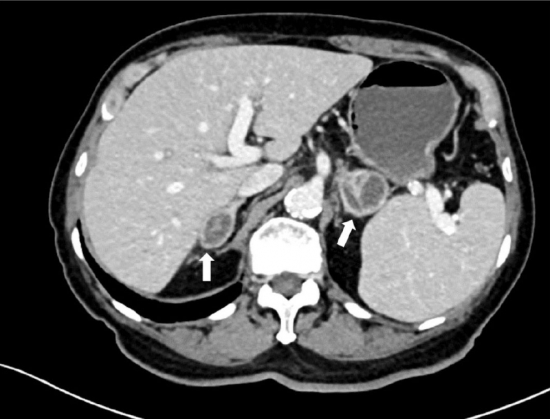
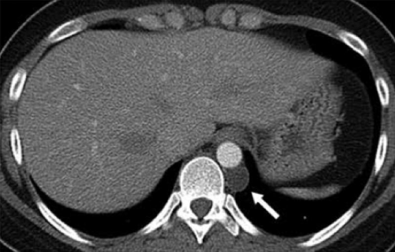
An 85-year-old man presented with a 3-month history of weight loss and anorexia. He had no history of tuberculosis and his physical examination and chest radiography were normal. Bilateral adrenal masses were seen on abdominal computed tomography scan. Serological test for HIV was negative.
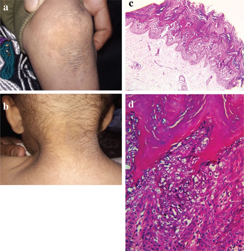
A 1.5-year-old Egyptian boy is presented with widespread, brownish, hyperkeratotic, and slightly verrucous plaques on the skin, since the age of two months. The patient has no history of blistering or generalized cutaneous redness at or after birth. Upon physical examination, brownish, verrucous plaques…
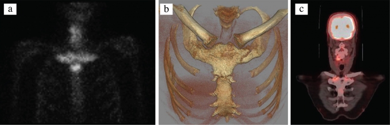
A 6-year-old male patient was presented with a two-month history of polyuria and polydipsia. The patient's 24-hour urine osmolality was 79 mOsm/kg and he lost 5.2% of his weight after 7-hour water deprivation test. His renal functions were normal, his polyuria and polydipsia resolved on treatment with 60 μg desmopressin…
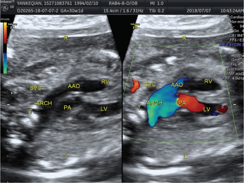
A 31-year-old singleton pregnant woman (gravida 2, para 1) was admitted to the Maternal–Fetal Medical Center, at 30 + 1 weeks of gestation, with a suspicion of fetal congenital heart disease. Prenatal echocardiography exhibited parallel initial segments of the aorta pulmonary arteries. The aorta originated from the right ventricle…
A 53-year-old male was evaluated for upper chest pain lasting three years. The pain was continuous, bilateral, progressive, and worse when the patient lay on his sides and during hyperabduction of the arms, radiating to the shoulders. Initially, it was responsive to non-steroidal anti-inflammatory drugs…
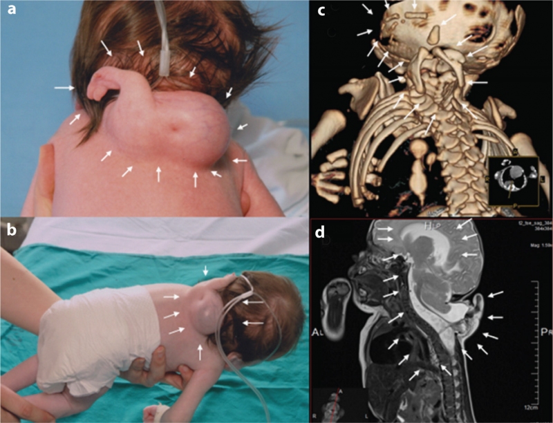
A female newborn infant was born with a dorsal mass on her cervicothoracic region. The mass consisted of accessory partial and hypoplastic upper limbs completely covered with skin. The dorsal mass measured approximately 10 cm in diameter, was soft, showed no movement, was sensitive to touch, and was associated with a spectrum of other anomalies…


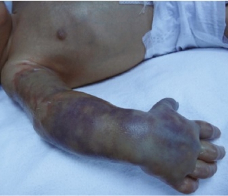
A 2190 g male neonate was born at 34 weeks of gestation by emergency cesarean section (fetal bradycardia). He admitted to the intensive care unit due to prematurity and perinatal asphyxia. He had tonic-clonic convulsions in the upper extremities on the second day of life refractory to ongoing phenobarbital, midazolam treatments. Phenytoin of 40 mg was intended to infuse over 30 minutes, through a peripheral venous cannula on the right hand. The purple-blue discoloration of the right hand and arm was observed at the 15th minute of infusion. The right arm had become edematous in hours.


Copyright © 2026 - Balkan Medical Journal